Interventional Management of Ectopic Ureters: A New Treatment Option
|
Ectopic ureters are the most common cause of urinary incontinence in juvenile female dogs, if you need more information about exactly what they are then please see my owner guide here.
A number of surgical methods have been described to correct this condition, however they are associated with a reasonably high complication rate and are, on the whole, relatively invasive procedures. For that reason, less invasive methods have been investigated for management of ectopic ureters, we currently use a technique called cystoscopic laser ablation in appropriate cases. This uses a combination of cystoscopy and, often, fluoroscopy to guide a very small ureteral catheter into the ectopic ureter, and then uses a laser fibre through the working channel of the cystoscope to ablate the wall of the ureter. This essentially moves the opening from the urethra to the bladder (or as close to the bladder as possible). Procedure:
Is the procedure suitable for all ectopic ureters? No, this procedure is only suitable for intramural ectopic ureters as it ablates the wall of the ureter as it travels through the urethra. The good news is that 95% of ectopic ureters are intramural so it is appropriate in most cases. The exact path of the ureter is charted before the procedure using x-rays, fluoroscopy, CT or cystoscopy. What equipment is required for the technique? This technique is usually performed by somebody experienced in cystoscopy, as it uses a rigid cystoscope to access the urethra. Ideally fluoroscopy is also used to assess the ureter and the location of the cystoscope. Lastly a surgical laser is required with an endoscopic laser fibre, we usually use a diode laser but Holmium:Yag lasers are also described. What is the recovery time? Most dogs are treated as a day case or stay for one night in the hospital, pain relief is routinely given for a few days and some dogs may show some signs of mild urinary discomfort. We also commonly instil topical local anaesthetic into the urethra. Will it fix the incontinence? As many dogs with ectopic ureters can have other concurrent urinary tract abnormalities, incontinence can remain in some cases. In one study around 50% of cases were continent with this procedure alone and the others required additional treatment (most commonly medical treatment of urethral sphincter mechanism incompetence). What are the potential complications? When performed by an experienced operator this is a very safe technique with a very low rate of complication. As with all interventional urology there is a small risk of damage or perforation of the urinary tract. More specifically some dogs do get a proliferative reaction at the site of laser ablation which can cause polyp/tissue formation and then cause additional problems, this is reported in around 3% of cases. Can the procedure be performed in male dogs or cats? The technique is most commonly performed in female dogs and is most straightforward in this setting. In male dogs the procedure can be performed with a flexible cystoscope (as long as the patient is large enough) or via an approach into the urethra through the perineum, which is therefore slightly more invasive. It may be possible in some cats, however this can be limited by size, as not all cats are amenable to rigid cystoscopy. |
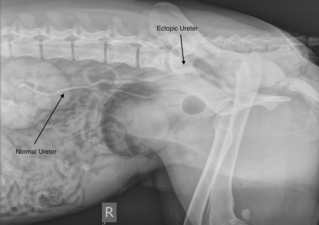
A contrast radiograph of a dog with an ectopic ureter
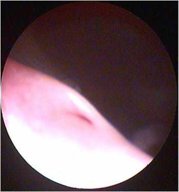
A cystoscopic image of a normal ureteral opening
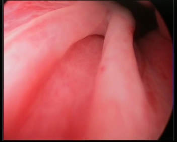
A cystoscopic image of an ectopic ureter
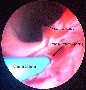
A cystoscopic image of an ectopic ureter
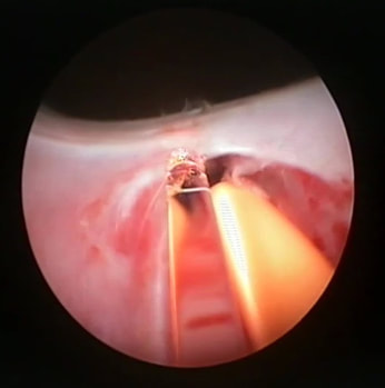
Laser ablation in progress
|
If you have any questions about this procedure, please feel free to contact me through the site.
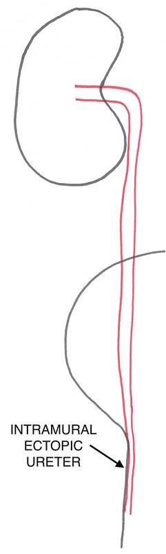
A diagram of an intramural ectopic ureter before treatment
|
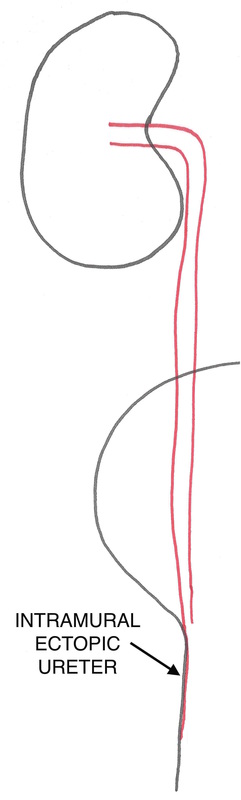
A diagram of an intramural ectopic ureter after treatment, note that the opening of the ureter has, effectively, been moved back to the bladder neck by ablation of the medial ureteral wall
|
References:
Veterinary Image-Guided Interventions. Edited by Weisse & Berent. Wiley Blackwell 2015
Evaluation of cystoscopic-guided laser ablation of intramural ectopic ureters in female dogs. Berent, Weisse, Mayhew, Todd, Wright & Bagley. JAVMA 2012, 240 (6), 716 - 725.
Use of cystoscopic-guided laser ablation for treatment of intramural ureteral ectopia in male dogs: four cases (2006-2007). Berent, Mayhew & Porat-Mosenco. JAVMA 2008, 232 (7), 1026 - 1034.